


Home to the Most Effective Distal Radial Fracture

Distal Radial Fractures Are Serious Injuries That Require Reliable, Expert Care
Wrist fractures are common — and nondiscriminatory. Even individuals with excellent bone health may experience a broken wrist if a traumatic blow or an unusual fall occurs. Alternatively, those with poor bone health (especially patients with osteoporosis) need only a minor fall or light blow to experience a break in the wrist.
In fact, fractures of the distal radius occur more frequently than any other bone in the arm. If you have experienced a wrist injury and think it may be a distal radial fracture, the distal radial fracture surgeons at Rothman Orthopaedic Institute can provide you with a diagnosis and a treatment plan.
No matter your age or state of your general orthopaedic health, pursuing specialized care following a distal radial fracture is paramount. Whatever type of subsequent treatment is recommended for your injury, the Hand and Wrist specialists at Rothman Orthopaedic Institute will ensure that you receive the most advanced, effective care available. Learn more below.
Distal Radius Anatomy
Within the forearm, there are two bones that support the rotation and pivot movements of the hand: the ulna and the radius. The ulna runs along the outside of the wrist, while the radius is situated along the inner side with closer proximity to the thumb. The radius is a shorter bone than the ulna but is wider in thickness at the wrist.
Types of Distal Radial Fractures
Fractures of the radius commonly occur approximately one inch from the bottom of the bone closest to the wrist. There is a multitude of positions in which the bone can break, however. Listed below are some of the different types of radial fractures which patients may experience.
-
Colles Fracture
The majority of fractures are characterized by a distinct upward tilt of the fractured distal radius end. The medical name for this type of distal radial injury is the Colles fracture. -
Intra-articular and Extra-articular Fractures
These types of fractures are less common than the Colles fracture. Intra-articular fractures occur when the fractured section of the radius extends into the wrist joint. Extra-articular fractures, meanwhile, do not extend into the wrist joint. -
Open Fracture
Open fractures are the most serious variety of distal radius fracture. This type of injury occurs when the fractured radial bone penetrates the surface of the skin. If you have experienced an open fracture, it is critical that you undergo immediate medical treatment to avoid serious infection. -
Comminuted Fracture
The comminuted fracture is another severe type of distal radius fracture. When a comminuted fracture occurs, the affected bone is broken into multiple pieces. The bone fragments may stay in relative alignment or become displaced.
Your Rothman Orthopaedic Institute Hand & Wrist specialist will classify your injury in order to recommend an appropriate treatment plan. In each of these fracture types, the grade of severity can vary based on a number of factors. Displacement of bone fragments, the exact location of the break, and the size, shape, and number of fragments will all be taken into account by your doctor. Additionally, your age, level of activity, general health, and any previous conditions will factor into which treatments are ideally suited for you.
Seeking Treatment
If deformity after an injury is obvious, the pain is excruciating and numbing, and the fingers are turning an abnormal color, visit a doctor as soon as possible.
It is strongly recommended that all patients seek medical care promptly following a fracture or suspected fracture. If you cannot undergo treatment on the same day as your injury, you are advised to immobilize your injured arm (with a splint, if possible) and wrist, ice the area, and keep the wrist elevated.
Before attempting these temporary treatment measures, speak on the phone with a doctor who can guide you through implementing at-home care correctly. Rothman Orthopaedic Institute physicians encourage patients to call whenever you are uncertain and have questions about at-home treatment and recovery.
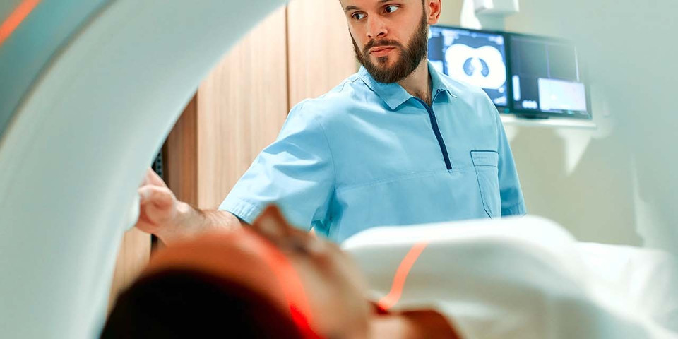
Diagnostic Examination at Rothman Orthopaedic Institute
No matter what the particulars of your injury may be, a medical examination by a qualified specialist is imperative. A physical examination at Rothman Orthopaedic Institute will not only assess the nature and severity of your injury but will also allow your doctor to determine the best steps forward for treatment, healing, and recovery.
What should you expect from your wrist examination?
-
First, your doctor will ask you to describe how your injury occurred. She or he will also use this time to inquire about your medical history (especially orthopaedic bone conditions, such as osteoporosis).
-
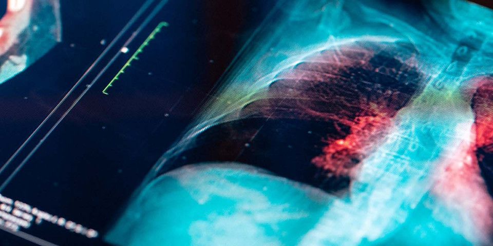
Then, your doctor will order x-rays. At Rothman Orthopaedic Institute, you will have on-site access to advanced x-ray diagnostic technology.
-
Finally, your doctor will assess the x-ray images of your injured wrist to definitively determine if a fracture has occurred. The location of the fracture, number of fragments, and any displacement or gaps between fragments will be noted.
At the conclusion of your examination, your doctor will discuss treatment options with you and offer expert recommendations.
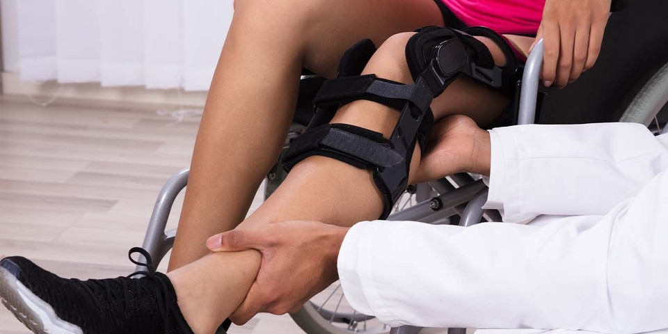
Distal Radial Fracture Treatment: What To Expect
Following your diagnostic examination, your distal radial fracture surgeon or physician will recommend the appropriate treatment method to align the fractured bone and stabilize the position of the radius and wrist. This may include non-operative realignment, splinting, casting, or surgical options.
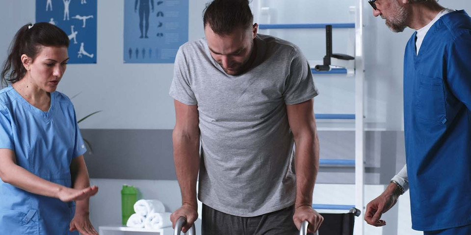
Your doctor will closely supervise the initial treatment and your body’s reaction to the prescribed method. Sometimes, casting or surgical dressing may need to be either loosened or tightened, depending on your needs. Distal radial fracture surgeons understand the individualized nature of the injury and seek to provide you with the customized treatment your injury demands.
The specifics of your treatment and recovery process will be determined by the type of fracture which you have experienced and, consequently, the type of treatment methods you have been prescribed. In the following section, we’ll take a closer look at the treatments that will typically be recommended for each type of distal radial fracture injury.
What Type of Distal Radial Fracture Treatment Do I Need?
Does your distal radius fracture require surgical intervention, or will non-operative treatment be sufficient? What types of surgical operations are recommended?
The answers to these questions will be determined to a great extent by the type of fracture you have experienced. Listed below are the typically recommended treatments for each type of distal radius fracture.
-
Colles Fracture Treatment
If you have experienced a Colles fracture that does not involve displacement of the fractured bone, your physician will usually advise non-operative treatment. This will involve splinting or casting to immobilize the affected wrist and enable healing.
In some cases, bones with minor displacement can be realigned without surgery through a process called closed reduction. If the bone fragments are seriously displaced, surgical “open” reduction through an incision will be needed. After surgery, casting or splinting will be applied. -
Intra-articular and Extra-articular Fracture Treatment
The recommended treatment methods for intra-articular and extra-articular fractures are comparable to those of the Colles fracture: closed reduction and casting or splinting for fractures in relatively good alignment and surgical reduction for displaced fragments. -
Open Fracture Treatment
Open fractures lend a special urgency to the treatment process as tissue exposure may lead to infection. This means that medical treatment will need to be undergone promptly (one hour to eight hours from the point of injury).
Open fractures always require surgical intervention. The surgical treatment process will involve cleaning and, in some cases, antibiotic treatment of exposed bone and tissue, followed by surgical reduction. External or internal fixation methods (such as plates and screws) will then be applied to maintain the correct structure and alignment of the bones. Following surgery, casting or splinting will be applied. -
Comminuted Fracture Treatment
The multiple fragments resulting from a comminuted fracture will usually require surgery (followed by casting or splinting) in order to heal properly, though closed reduction and immobilization may be sufficient in certain cases.
It is advised to update your doctor within 24 hours of surgery to confirm that swelling has gone down, pain has lessened, and finger movement is possible. Whichever treatment you ultimately undergo, your Rothman Orthopaedic Institute wrist specialist will work closely with you, guide you through the recovery stage, and ensure the long-term success of your treatment.
Schedule an Appointment With a Distal Radial Fracture Surgeon
Rothman Orthopaedic Institute provides comprehensive care with cutting-edge technology. As true clinical specialists, our distal radial fracture surgeons have the education and experience needed to provide exceptional, effective care to patients of every age and health history.
Our Hand and Wrist surgeons have all received sub-specialized training. This means Rothman Orthopaedic Institute patients have ready access to ideally-equipped physicians who are the best in their field.
For more information or to schedule an appointment, please visit us here or contact us at 1-800-321-9999.
Related Specialties
Related Physicians

Related Conditions
Related Treatments
Related Services
Related Programs
-

Cartilage Restoration Institute
This is a center where patients can go to have their disabled joint biological resurfaced, realigned, and stabilized without having the joint replaced by artificial materials such as metal and plastic. It is well known that the outcomes of patients under the age of 50 undergoing artificial joint replacement are not as good as we would like. Therefore we feel the future of Orthopaedics is to try to restore a joint back to its original anatomy by realignment, ligament reconstruction, and cartilage restoration.Read More